- "Manavdaya Trust Sankul", Saroli, Dist. Surat, Gujarat.
- OPD : Mon - Sat 10.00 - 06.00. Sunday CLOSED
-
 24 Hour Helpline
-
 Emergency
-
 Ambulance
Radiology
Radiology Department

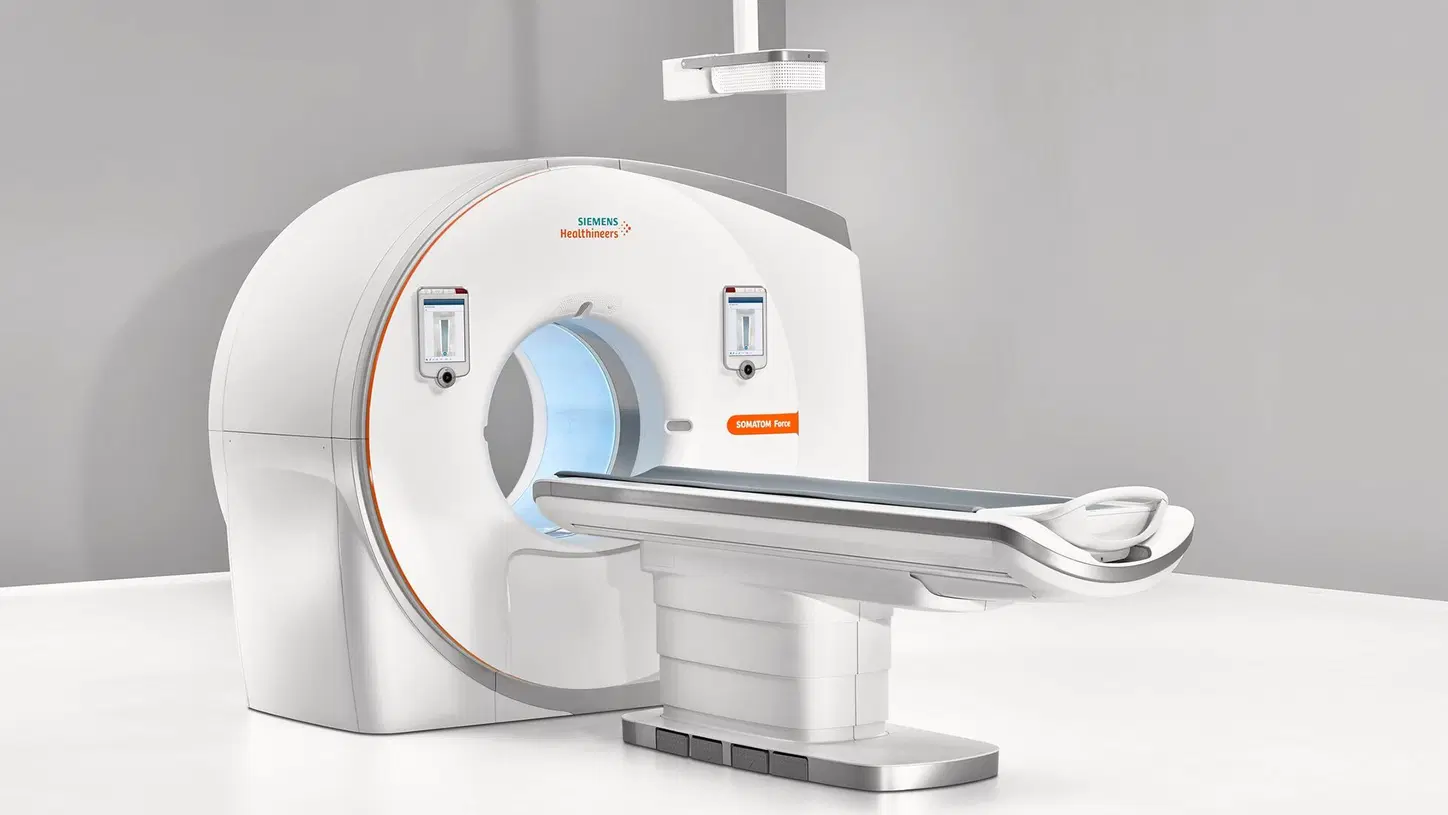
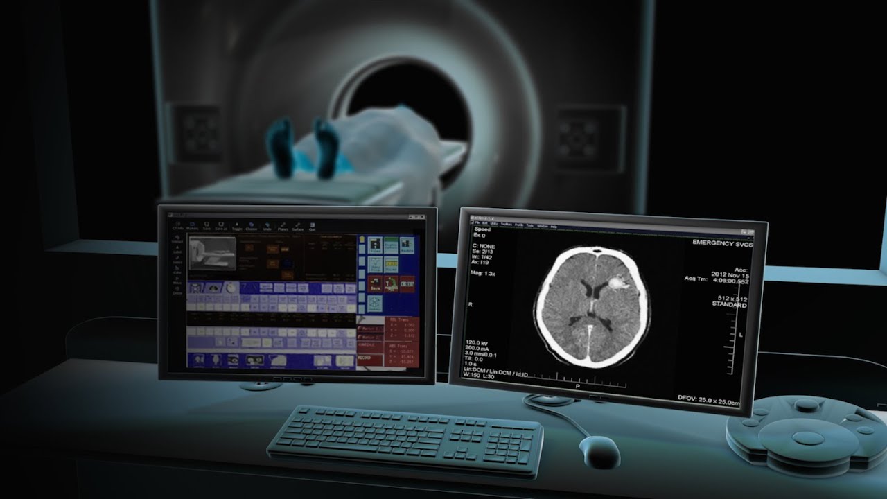
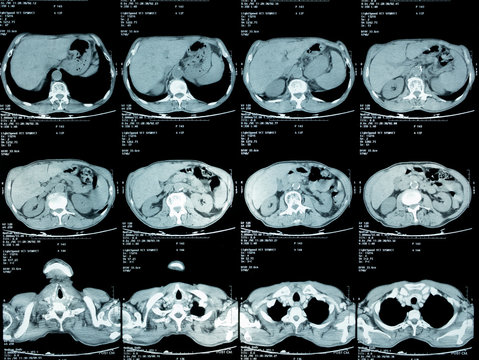
256 Slice CT ScannerThe 256 Slice CT Scanner is the ultimate CT solution for body imaging. It fuses the latest and fastest multiline technology with dedicated clinical applications delivering unprecedented image quality. |
All parts of the body can be scanned with CT. For each part, there are specific indications or reasons for scanning. Your referring doctor and the consultant radiologist are the best judges of the usefulness of CT scanning in a specific situation and areas.


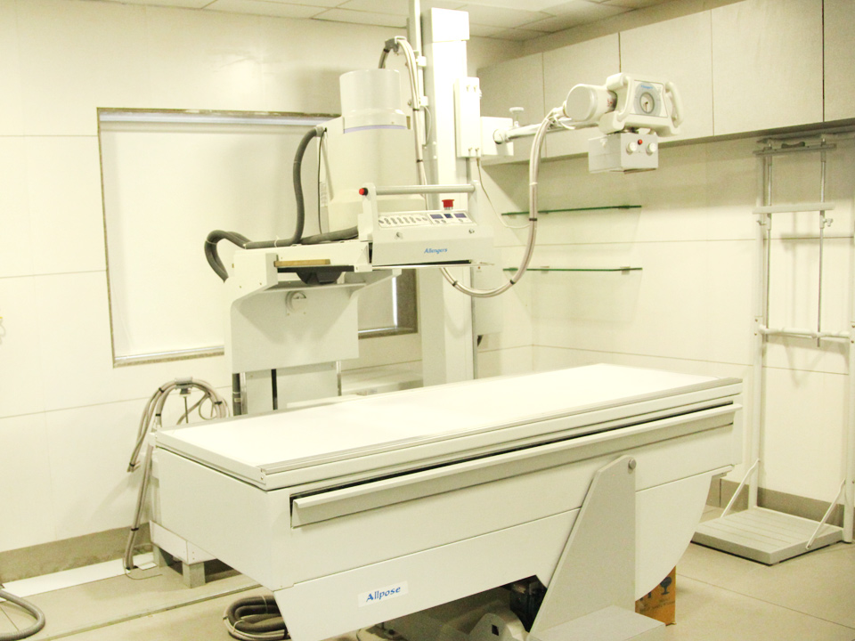
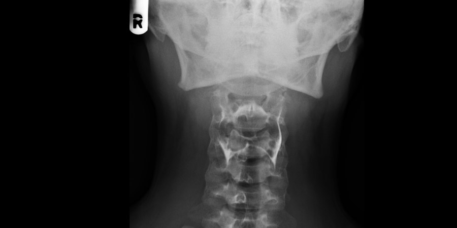
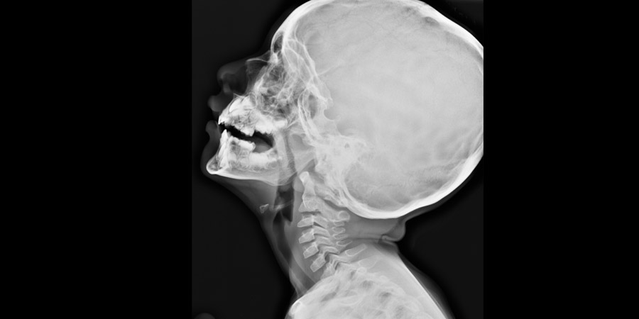
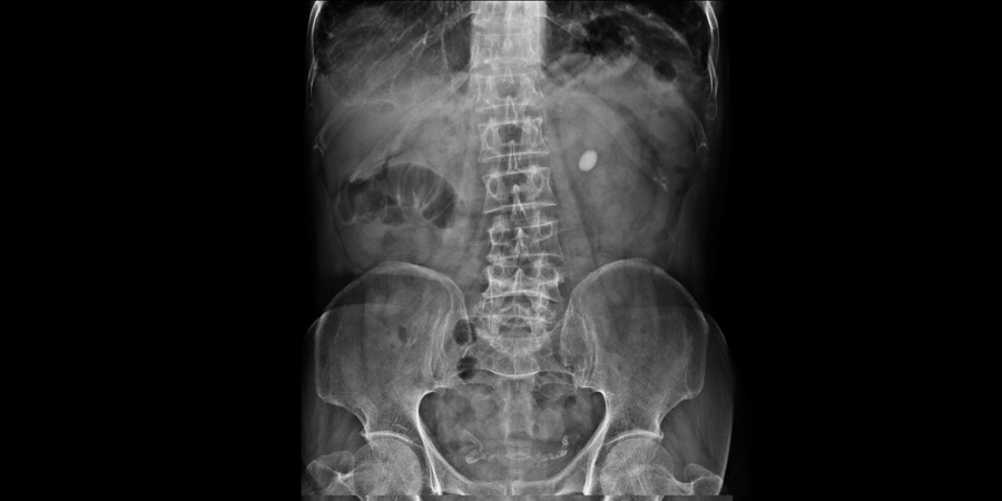
Digital X-rayDigital radiography systems from Bharat Cancer Hospital are as individual as your X-ray routine, cover virtually all clinical applications, and offer you workflow optimization, imaging excellence and investment confidence. |
All images obtained using digital systems are called digital X-rays. The common digital method used is the CR system that produces digital X-rays on a computer. See the accompanying picture for an idea of how this works.
Types of X-Ray Exams:
- Chest
- Shoulder, Arm or Hand
- Hip, Leg or Foot
- Spine
- Sinus
- Skull
Types of X-ray Procedures:
- HSG
- IVP
- MCUG
- RGU
- BMFT
- Loopogram

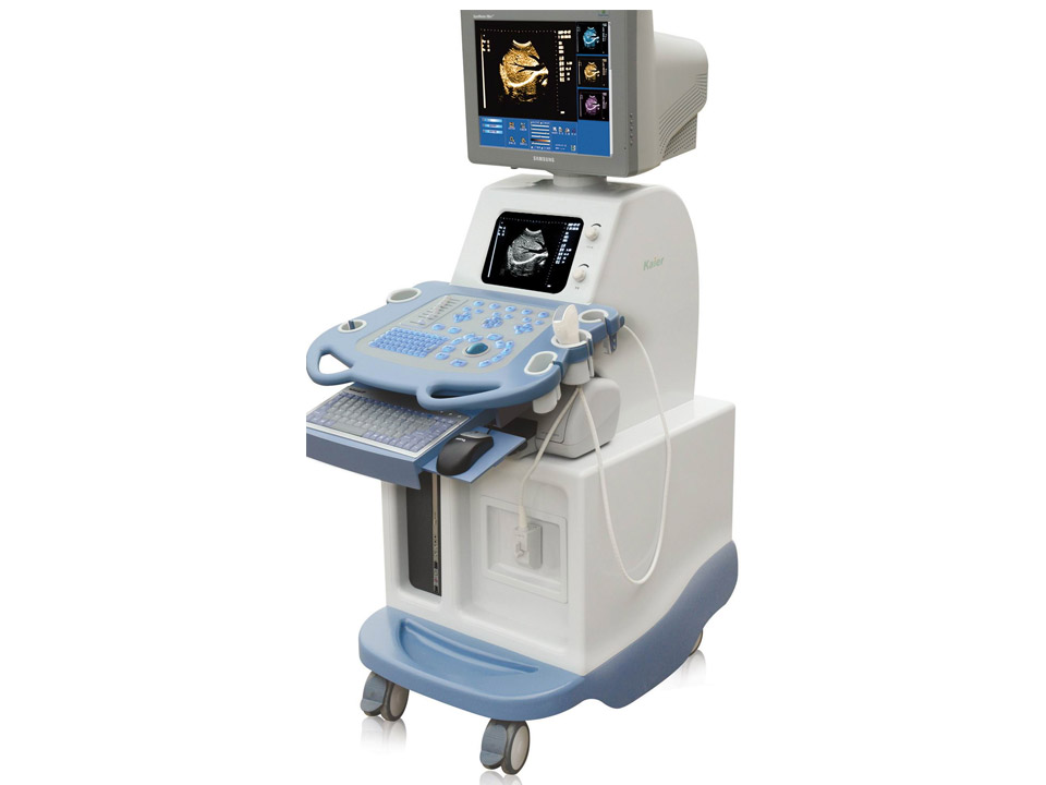
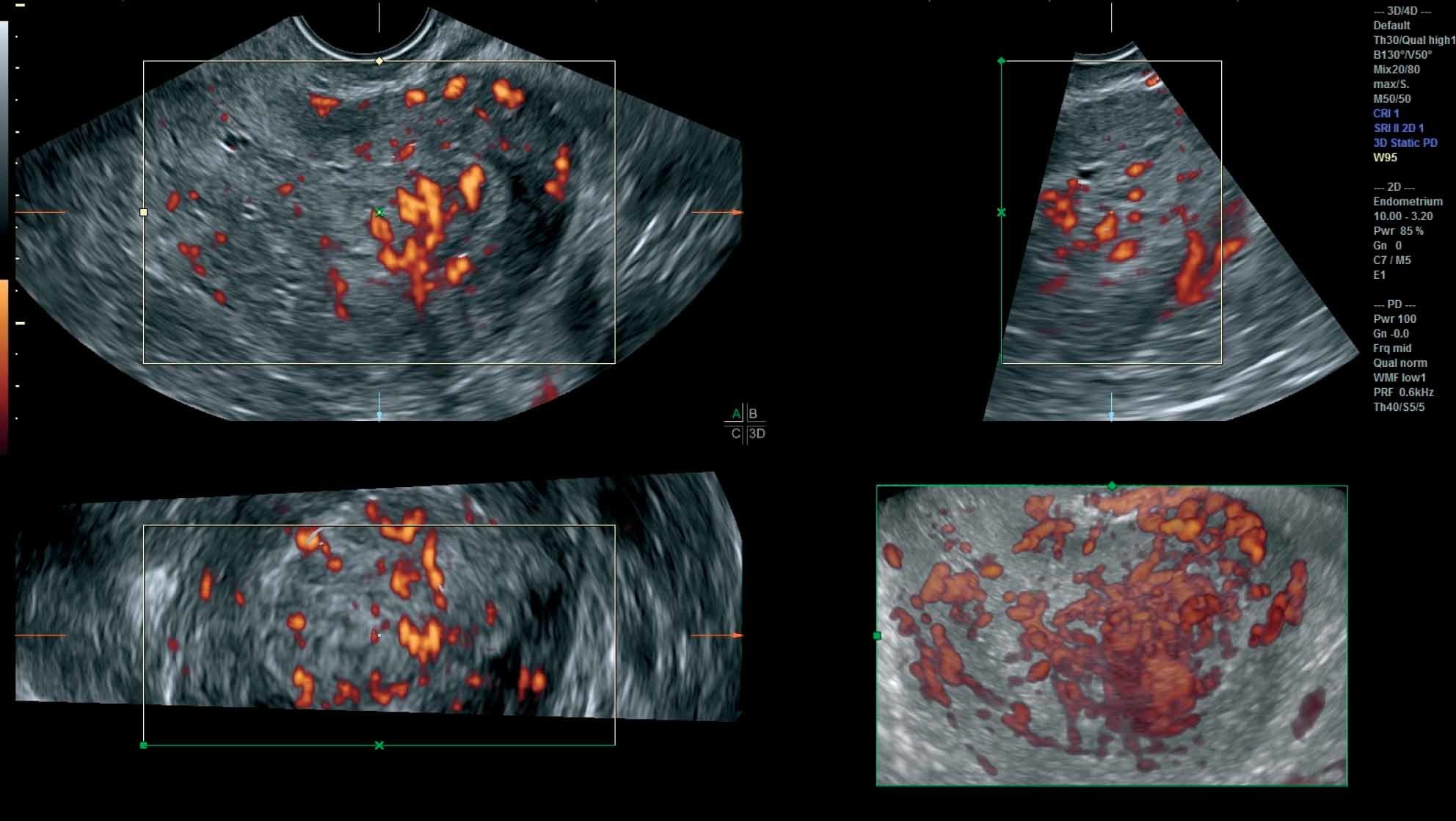
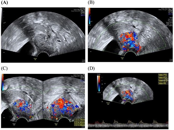
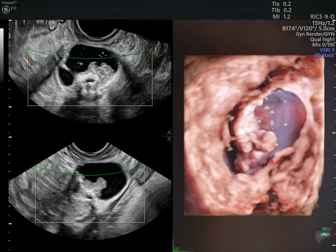
3D/4D Sonography3D scans show still pictures of cancer treatment in three dimensions. 4D scans show 3D images of cancer treatment, with time being the fourth dimension. |
3D and 4D scans are considered as safe as 2D scans, because the images are made up of sections of two-dimensional images converted into a picture. However, experts do not recommend having 3D or 4D scans purely for a souvenir photo or recording, because it means that you are exposing your baby to more ultrasound than is medically necessary. Some private ultrasounds can be as long as 45 minutes to an hour, which may be longer than recommended safety limits.
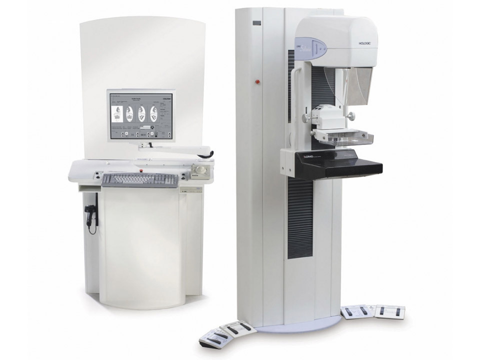
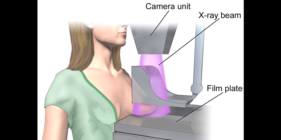
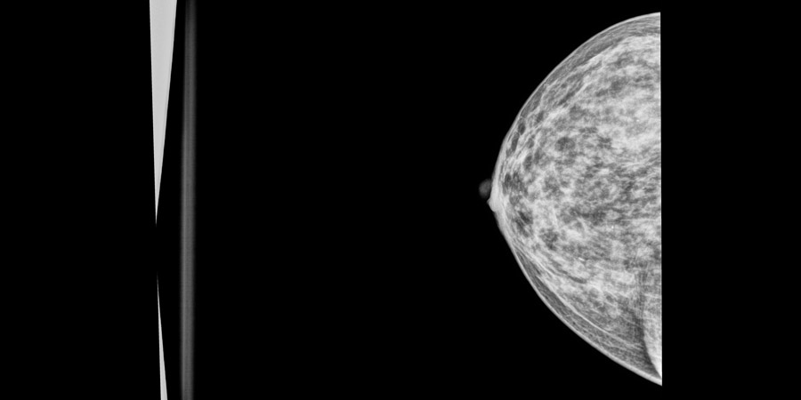
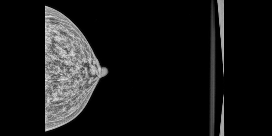
MammographyDiagnostic Mammography can sometimes be required following a screening mammography. The goal of diagnostic mammography is to pinpoint the exact size and location of breast abnormality and to image the surrounding tissue and lymph nodes. Sometimes additional views, ultrasound, or fine wire maybe required if the radiologist would like to focus in on a specific area, or if results are obscured, ie. high density breast tissue. The radiologist may recommend that a patient return for a follow-up mammogram, typically in six months. |
You will be asked to undress from the waist up so it is advisable to wear comfortable cloths. Do not wear underarm deodorant, powders, ointments, or creams on your breasts on the day of the exam. These can show up on the mammogram images.
There will be female Technicians as well as Radiologist to perform the mammography.


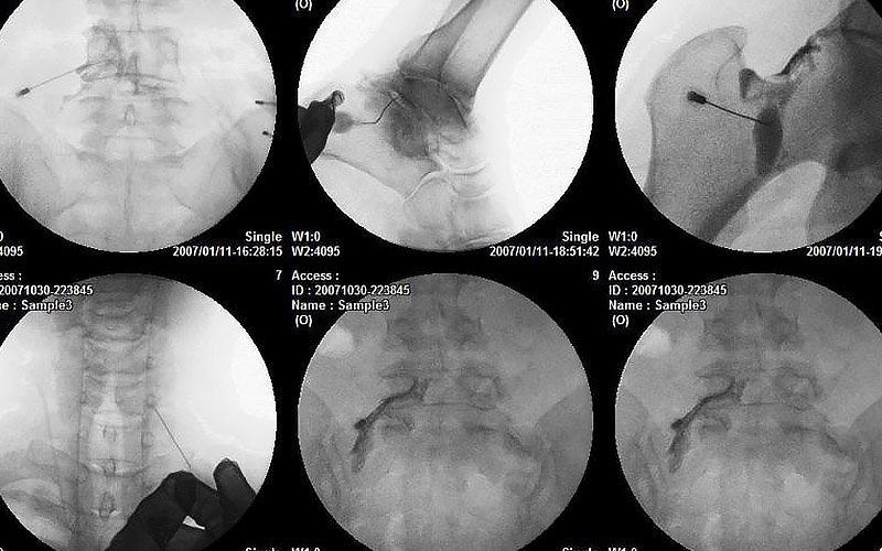
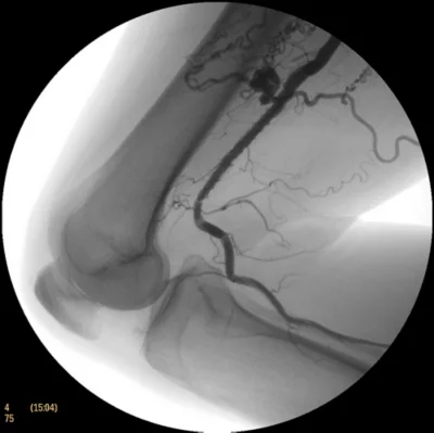
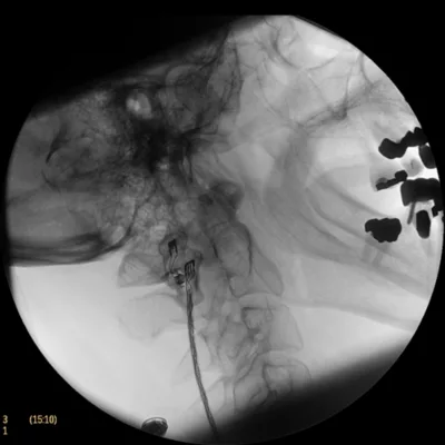
C-ARMTo assess the radiation dose reduction capabilities and the image quality of a new C-arm system in comparison to a standard C-arm system. |
Both cohorts showed no significant differences with regard to patient characteristics, tumor burden and fluoroscopy time. The new system resulted in a statistically significant reduction of cumulative DAP of 72% compared to the old platform (median 76 vs. 269 Gy*cm2). Individually, Fluoro-DAP and DSA-DAP decreased by 48% and 77% (p = 0.012 and p < 0.01), respectively. No statistically significant differences in DSA image quality were found between the two imaging platforms.